CUSTOMER SUPPORT
Building long-term partnership with our customers is our commitment in this industry. At EverBio, we understand that we need to deliver more than just a functioning equipment to meet customer’s criteria. The core value of EverBio’s equipment is far more than just the technology that puts the components together. Durability and flexibility is embedded in the spirit of the machine when we first craft them from the sketch board.

Attempts have been made to automate the process of tissue microarray construction.
Various machines are now available (automated tissue arrayer) which may array single and multiple tissue microarray blocks in an even shorter period of time than that performed manually.
Furthermore, digital techniques have taken center stage in clinical medicine as well as in biomedical research. Recently pathology has begun to embrace this digital revolution. The first development was a high throughput microscopic slide scanner that can convert traditional glass microscopic slides into digital images that can be stored, retrieved, shared through the internet and, most importantly, algorithmically analyzed. The other development was the creation of algorithms that can identify and quantitate immunohistochemical staining patterns and specific histological features, such as nuclear size and mitosis.
Such an algorithm can be applied to digital images of microarray. Digital pathology and imaging technology that can scan tissue microarrays into “virtual slides” with high resolution and that can analyze their images algorithmically will accelerate the discovery of new predictive biomarkers.
MALDI MSI (matrix-assisted laser desorption/ionisation) is a molecular imaging technique which does not require previous knowledge of analytes. It is therefore suitable for the discovery of new biomarkers. However, the success of biomarker research depends substantially on the analysis of large numbers of well-characterized samples. This is usually limited by the availability of such samples and, equally important, time. The analysis of tissue microarrays (TMAs) with MSI can overcome these limitations because tissue microarrays allow for the analysis of hundreds of specimens in a single experiment under identical analytic conditions.
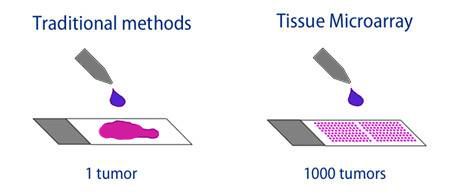
A tissue microarray is a collection of small tissue cores (usually with a diameter of 0.6-1.0 mm) taken from donator tissue blocks and transferred into an empty paraffin block. This allows for the study of tissue sample taken from up to 1000 different individuals in one experiment.

Tissue microarrays can either be built from formalin-fixed paraffin-embedded (FFPE) or from fresh-frozen tissue material. Both kinds of sample material can be used for analysis by mass spectrometric imaging.
Study of large-scale tissue microarrays
Large-scale tissue microarrays with formalin-fixed paraffin-embedded tissue cores can hold up to 1000 samples of different tumors per block. With mass spectrometric imaging, an untargeted high-throughput analysis of a huge number of samples can be performed under identical analytic conditions.
Since the large-scale tissue microarrays contain FFPE tissue cores, specific sample preparation methods are required prior to mass spectrometric analysis, including heat and acidic or basic pretreatment and on-tissue tryptic digestion.
Histology is the study of the microscopic structure of tissues. The study of pathological cells is known as Histopathology. By identifying morphological changes in tissues, it is possible to see characteristics of certain diseases.
Steps in Tissue Processing:
- Fixation
- Embedding
- Dehydration
- Clearing
- Sectioning
- Mounting
- Dewaxing
Steps in Tissue Processing:
- TMA Processing
- Sectioning
- Mounting
- Dewaxing
Fixation
-To preserve tissue architecture, tissue must first be fixed.
-Chemical reagents are used to hold tissue components in their normal places during the subsequent processing.
-They also prevent tissue from rotting and auto-digestion by leaked lysosomes.
-Fixatives have binding sites to form cross links between adjacent tissue proteins (this is how they preserve tissue).
-Microbes bind to spare binding sites and are deactivated, this is how tissues are protected from microbial attach.
-Fixative only proseve proteins.
Embedding
-Embedding tissues provides them with physical support so tissue can be sectioned thinly.
-The process involves hardening the tissues by replacing the water with resin or wax.
-Neither was nor resin are miscible with water however, so water is gradually replaced with alcohol in the process called dehydration.
-Once tissue has been dehydrated, it can then go through a process called clearing.
-As wax and resin are not miscible with alcohol either, the alcohol is replaced with a organic substance called xylene and the tissue goes transparent.
-Finally the solvent is replaced with hot molten wax (58.C) with is cooled to harden.
-The dehydration process can damage and alter tissue.
-The clearing process dissolves alcohol and lipids as well.
